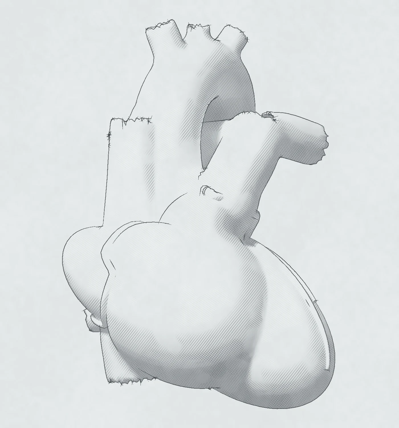
Almost every echocardiographic report includes Doppler calculations of flow and pressure. The same flow passes every level of the outflow: where the area falls, the velocity rises and the pressure falls. RV systolic pressure is 4 × (peak TR velocity)² plus the RA pressure estimated from the IVC.
Flow is the same at every level
flow rate = area × velocity · A₁ × V₁ = A₂ × V₂
Blood does not compress, so in each instant the same flow passes every level of the outflow: the LV outflow tract, the aortic valve and the aorta. The flow rate through an orifice equals the area of the orifice multiplied by the velocity of the blood in it. Where the area is small, the velocity is high: an orifice with a quarter of the area carries the same flow at four times the velocity. For a given orifice area, the velocity and the gradient increase with the flow rate and decrease when it falls.
The volume that passes in one beat equals the area multiplied by the VTI, and it is the same at each level: the stroke volume ejected through the LV outflow tract all passes through the aortic valve. The continuity equation uses this: the aortic valve area equals the LVOT area multiplied by the LVOT VTI and divided by the aortic VTI. Chapter 12 calculates the valve area in this way. The velocity ratio, the LVOT velocity divided by the aortic velocity, gives the valve area as a fraction of the LVOT area without the LVOT diameter. It approaches 1 without stenosis, and a ratio of 0.25 or less suggests severe stenosis.
Stroke volume from the LVOT
Stroke volume is the LVOT area multiplied by the LVOT VTI. The LV outflow tract is assumed to be circular, so its area is 0.785 × d², with the diameter d in cm. The LVOT diameter is measured in a zoomed parasternal long-axis view from inner edge to inner edge in mid-systole. The LVOT velocity is recorded with pulsed-wave Doppler from the apical five-chamber or apical long-axis view, with the sample volume just below the aortic valve. Both measurements should be made at the same level.
Because the diameter is squared, a small error in its measurement produces a large error in the calculated stroke volume. Repeated LVOT diameter measurements vary by 5 to 8%, which is the largest source of error in the continuity equation. A 2 mm error in the diameter of a 2.0 cm outflow tract changes the calculated stroke volume by approximately 20%.
Normal resting values from catheterization studies are a stroke volume of 60 to 100 mL, a cardiac output of 4 to 8 L/min and a cardiac index of 2.5 to 4.0 L/min/m².
Pressure falls where velocity rises
ΔP = 4 (V₂² − V₁²) ≈ 4 V₂²
As the blood speeds up into a narrow orifice, pressure is converted into velocity, and the pressure is lowest at the vena contracta, the narrowest part of the jet. The simplified Bernoulli equation calculates the pressure difference in mmHg from the velocity in m/s. It assumes that viscous losses and acceleration are negligible and that the velocity proximal to the narrowing can be ignored. When the proximal velocity is over 1.5 m/s, or the jet velocity is under 3.0 m/s, the proximal velocity is included: ΔP = 4 (V₂² − V₁²).
Past the orifice, the jet slows in the aorta. Part of its kinetic energy is lost as heat in turbulence, and the rest is converted back into pressure, which rises again: this is pressure recovery. The recovered pressure equals 4V² × 2 EOA/AoA × (1 − EOA/AoA), where EOA is the effective orifice area and AoA is the area of the ascending aorta. Doppler measures the pressure drop from the LV to the vena contracta, while a catheter measures the net drop from the LV to the aorta. In most adults with aortic stenosis the recovery is small, but when the ascending aorta is narrower than 30 mm, the Doppler gradient can be significantly higher than the net gradient.
The maximum gradient is the maximum instantaneous pressure difference across the valve. The mean gradient is the average of the instantaneous gradients over ejection, and it cannot be calculated from the mean velocity.
Flow convergence (PISA)
Blood accelerates toward a regurgitant orifice in roughly hemispheric shells. Color Doppler shows the shell at the aliasing velocity (chapter 03), and the flow rate through the orifice is the area of that shell × the aliasing velocity. Chapter 15 derives the effective regurgitant orifice area (EROA) and the regurgitant volume from it, and chapter 16 applies it to TR.
The vena contracta of a regurgitant jet is its narrowest part, at or just downstream of the orifice. It is slightly smaller than the anatomic orifice, and its cross-sectional area is a measure of the EROA. The regurgitant fraction is the regurgitant volume divided by the stroke volume through the regurgitant valve.
Pressure gradients and RV systolic pressure
RVSP = 4 × (peak TR velocity)² + RA pressure
The peak TR velocity gives the pressure difference between the RV and the RA, and the RA pressure estimated from the IVC is added to obtain the RV systolic pressure.
Without RV outflow obstruction, pulmonic stenosis or proximal PA stenosis, the RV systolic pressure equals the PA systolic pressure. In significant TR, the RV and RA pressures can equalize early; the CW signal then becomes triangular and underestimates the RV–RA gradient (chapter 16). A faint TR signal also underestimates it.
A resting peak TR velocity of 2.9 m/s or more, or of 2.8 m/s or more with two other echocardiographic signs, suggests pulmonary hypertension. An IVC wider than 2.1 cm is one of those signs. Chapter 13 shows the RV's response to a pressure load: its size, wall, function and septum.
RA pressure from the IVC
In the subcostal long axis, the caliper crosses the IVC perpendicular to its long axis, 1 to 2 cm from the RA, at end-expiration, where the diameter is usually largest. A second recording follows the IVC through a sniff. The collapse is the fall in diameter, as a percentage of the expiratory diameter. M-mode measures the diameter and its change with breathing more accurately than 2D imaging. Its line must cross the IVC at a right angle: an oblique line overestimates the diameter.
The 2025 ASE guideline gives the RA pressure as one of three values:
- 3 mmHg (range 0–5): IVC ≤2.1 cm with ≥50% collapse.
- 8 mmHg (range 5–10): IVC ≤2.1 cm with <50% collapse, or IVC >2.1 cm with ≥50% collapse.
- 15 mmHg (range 10–20): IVC >2.1 cm with <50% collapse.
The two mixed patterns are indeterminate. Secondary signs of a raised RA pressure then decide: RA enlargement, an atrial septum that bulges into the LA throughout the cardiac cycle, a restrictive right-sided diastolic filling pattern, a tricuspid E/e′ ratio >6 and a hepatic vein systolic filling fraction <55%. With none of these signs, the estimate falls to 3 mmHg. With signs present, it rises to 15 mmHg. If uncertainty remains, the estimate is 8 mmHg.
A value of 20 mmHg can be considered when the IVC is wider than 2.5 cm and collapses less than 50%, with dilated hepatic veins. When the patient cannot sniff, the collapse with quiet breathing replaces the sniff, with a cut-off of 20%.
Normally, hepatic vein S is larger than D. A raised RA pressure lowers S/D below 1, and a systolic filling fraction, S/(S + D), below 55% is a sensitive and specific sign of it. In severe TR, S reverses (chapter 16).
The IVC runs on the right side of the spine, within the liver, and the hepatic veins drain into it. The aorta lies in the midline, apart from the liver, and does not change with respiration.
Pitfalls of the IVC
- Positive-pressure ventilation. The collapse of the IVC does not estimate the RA pressure. If the RA pressure is clinically relevant, an invasive measurement should replace the estimate. Secondary signs can show whether it is normal or raised. If uncertainty remains, the estimate is 8 mmHg.
- Athletes, young adults and pregnancy. A dilated IVC can be normal in athletes and healthy young adults and is common in pregnancy.
- Raised intra-abdominal pressure. It can collapse the IVC despite a raised RA pressure.
Key points
- The same flow passes every level, so where the area falls, the velocity rises and the pressure falls: ΔP = 4 (V₂² − V₁²), simplified to 4V₂² when the proximal velocity is small.
- Past the valve, part of the pressure returns as the jet slows (pressure recovery); with an ascending aorta under 30 mm, the Doppler gradient can exceed the net gradient.
- Stroke volume equals the LVOT area × the LVOT VTI, and an error in the LVOT diameter is squared in the calculation.
- RV systolic pressure equals 4 × (peak TR velocity)² plus the RA pressure estimated from the IVC.
- The IVC, measured 1 to 2 cm from the RA at end-expiration, gives 3 mmHg when it is ≤2.1 cm and collapses ≥50% with a sniff, 15 mmHg when it is >2.1 cm and collapses <50%, and 8 mmHg in the mixed patterns unless secondary signs decide.